Q. 11. Describe the structure and function of Bulbils in Cycas. (2006, 08, 11)
Ans. Bulbils: -
Bulbils are developed sometimes on the old stem of Cycas for vegetative reproduction. These are developed between the slits of permanent leaf bases on the basal portion of stem from parenchymatous cells of cortex e.g., Basal portion of each bulbil is surrounded with scale leaves and some foliage leaves are also present in the centre. Frequently these bulbils are produced in Cycas revoluta, because male plants of this species are not found in northern India and sexual reproduction does not oocur up to some extant. In this species the development of male and female bulbils occur on male and female plants separatley. After maturity bulbil fall down on earth and develops a new plant.

Q.12. Describe the species of Cycas in India. Where do they occur? (2006)
Ans. Species of Cycas in India and their Distribution: -
There are six species of Cycas occuring in India. Of them four are naturally produced and two species are cultivated in gardens. The distribution of naturally occuring species is as under:
1. Cycas circinalis : This species is found in decidous forests of South India, Orissa, Srilanka and also in the heavy rain fall area of the hill forests of mals of Puri and Angul.
2. Cycas pectinata : This species is widely distributed in the regions Sikkim, Someshwar hills of Bihar, Eastern Assam, Manipur, Chitgaon and East Bangal.
3. Cycas rumphii : Species of C. rumphii is mostly found in Andman - Nichobar and coastal region of Cokas island.
4. Cycas beddomei : This species is found in Chennai, Kerala, South India, Calicut, Eastern Andhra Pradesh. Beside these Cycas relvoluta (Sago Palm) and Cycas siamensis is cultivated in different gardens in India.
Q.13 Write short note on coralloid root of cycas. (2005, 07, 10, 12)
Related Questions -
Q. Describe the anatomy (internal structure) of Coralloid root of cycas and write the function of Coralloid root. (2013)
Ans. Coralloid Root of Cycas: -
T. S. of the coralloid root shows zone of cork followed by cork cambium.

(i) having poorly developed secondary tissues or none and
(ii) the cortex which is differentiated into three distinct zones :
(a) the outer cortex, composed of compact polygonal cells,
(b) the inner cortex of thin walled parenchymatous cells and
(c) the middle cortex forming the algal zone. Algal zone, which is usually one cell wide, consists of loosely connected thin walled and radially elongated cells and abundant large intercellular spaces. Intercellular spaces present in between these radially elongated cells are occupied by certain algae (Anabaena cycadacearum; Nostoc punctiforme and sometimes Oscillatoria and diatoms),some fungi and a few bacteria (Pseudomonas and Azotobacter). Each cell present in the algal zone contains a single nucleus and cytoplasm.
According to Schneider some nitrogen fixing bacteria and the blue green algae are present in the roots are symbiotic in nature. The algal zone serves two important functions - alvalian and nitrification. Zach (1910) is of the view that there is parasite relationship discarded the symbiotic relationship.
According to the observation of Schaede (1944) no bacteria are present in the corolloid roots. The mucus of the intercellular space of the roots provides the nourishment to the algae.
Q.14. Describe the mature ovule of cycas. (2008)
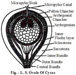
Ans. Mature Ovule: -

The ovule is erect. Each. ovule of the Cycas consists of a single massive integument which surrounds the mass of parenchymatous tissues the nucellus (it is diploid (2x) in nature) around except at the upper end where a narrow passage, the micropyle is formed. The main body of the ovule is the nucellus or Megasporangium proper. The integuments in a well developed ovule consists of three easily recognised layers, an outer and in inner fleshy layer of rather simple structures and a very complex. Stone layer between them. The vascular supply of the ovule is practically uniform. The outer set of vascular strands consisting of a few vascular bundles, traverse the outer fleshy layers integument and goes from the base of ovule to the micropyle and an inner set of vascular strands go the inner fleshy layer extending to the free portion of nucellus. The vascular strands are composed of measarch xylem. At the apex of the nucellus, a vigorous growth results in the formation of the characteristic beak, the nucellar beak, which is forced up into the micropyle. The centre of the beak becomes hollow the form the pollen chamber in which pollen grains or microspores collect after pollination. The pollen chamber is in communication with the micropyle. Deep within the nucellar tissue is seen a single large embryosac.