
Q.7 Describe in detail the life cycle of Marchantia. ( 2008 )
Related Questions -
Q. Draw a labelled diagrams of life cycle of Marchantia. (2013)
Q. Describe the gemma cup of, Marchantia. (2005, 06, 07, 11)
Q. Describe the sexual reproduction in Marchantia. (2007)
Q. Draw labelled diagram of:
(i) Capsule of Marchantia (2007)
(ii) V.S. of Marchantia antheridophore. (2006)
Ans. Occurence: -
The genus Marchantia represented by 65 species distributed all over the world In India only 10 - 12 species have been reported. All the species are terrestrial growing on moist shady places, damp soil, moist rocks, banks of water. The most common species is M. polymorpha, M. palmata, M. nepalensis and M. simlona.
Gametophytic phase: -
External features of Thallus: -

The plant body Marchantia is gametophytic or haploid in nature. The plant body is thallus like, prostrate, dorsoventrally differentiated, dichotomously branched, they posses a prominent midrib the thallus is dark green in colour. The thallus may attain a length of 5cm. to 10 cm. Each lobe of thallus possess a distinct midrib and an apical notch, its margin is weavy. The dorsal surface of thallus has small rhomoidal to polygonal areas called areole which marks the position of stoma like opening into the air chamber. On the ventral surface of thallus on either side of the midrib present two or more rows of scales which protect the growing point. On the lower side of central surface of thallus are present the rhizoids which are of two types smooth walled and tuberculated and do the function of fixation and absorption.

On the dorsal surface certain cup like structures, the gemma cup are present. These are special vegetative reproductive bodies.
In some cases certains special upright branches are developed at the growings apex. These branches bear the sex organs and are of two kinds, antheridiophores and archegoniophores.
Internal Structure of Thallus: -
The V. S. of Marchantia thallus shows two distinct regions:
(i) Upper Photosynthetic Region - The photosynthetic region is present towards upper surface of thallus.

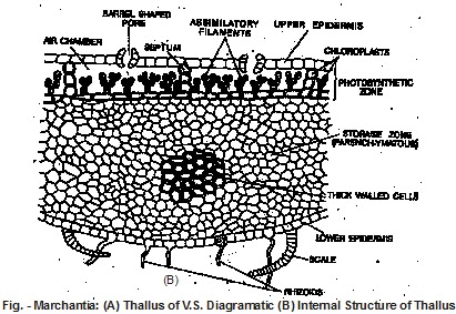
The photosynthetic region is distinguishble into photosynthetic air chembers which consist of photosynthetic filaments. Each cell of filament contains chloroplast. The photosynthetic filaments are meant for production of organic food.
(ii) Lower Storage Region: -
Just below the air chambers (photosynthetic region), there is compact colourless ventral region known as storage region. It contains parenchymatous cell which consists of starch as reserve food. This part is covered by single-layered lower epidermis towards ventral surface of thallus. The lower epidermis bears smooth walled and tuberculate rhizoids.
Reproduction: -
1. Vegetaitve Reproduction: - It takes place in favourable conditions by the following methods:
(i) By Death and Decay of Older parts : The older parts of the thallus decay and when it reaches up to dichotomously branched apices, they are separated. Each apex develops into a new plant.
(ii) By Adventitious Branches : In some species of Marchantia adventitious branches from the ventral surface of the thallus. These are separated from the parent plant and give rise to new thalli.
(iii) By Gemmae : In Marchantia, vegetative reproduction is mainly occurs by specialised reproductive bodies called gamme. Gemmae are formed in the gemma cup. These cups are developed on the dorsal surface along the midrib of the thallus.
Each mature gemma is multicellular, discoid body, which is vertically inserted in the gemma cup and attached to the floor with one-called hyaline stalk. It is 8 shaped in front view and looks biconvex lens-shaped from the side.

It is several cells thick in the middle and gradually thin towards the margin. Two growing points are situated in the shallow notches, one on each lateral margin, opposite to one another.
All the cells of gemma are green due to presence of chloroplasts, hyaline and large in size. These cells gives rise to rhizoids when the gemma germinates.
The gemmae get detached from their stalks in presence of water and forced out the cup they fall on the soil and germinate. The rhizoidal cells grow into the rhizoids. The marginal row of apical cells, situated at the lateral notches become activated and divide to produce young gametophytes, thus two young thalli are formed.
2. Sexual Reproduction: - In .Marchantia sexual reproducion is highly advanced oogamous type. The male sex organs are antheridia and female sex organs are archegonia which are produced on antheridoiphore and archegoniophore respectively. Marchantia is usually hetrothallic i.e. sex organs are borne on separate thalli.
Antheridiphore - It is having a cylindrical stalk about 1-3 cm in length and an 8 lobed disc. The internal structure of antheridiophare is similar to thallus. Each lobe of this disc is having a row of antheridia developed in acropetal succession. Each antheridium is present in antheridial chamber, which is a flask shaped structure and opens by a small pore on the upper side of disc. Each antheridium produces a large number of uninucleate, tubular biflagellate antherozoids.
Development of Antheridium - The antheridum develops from a superficial cell called antheridial initial, this antheridial initial increases in size and divides transversely into upper outer cell (primary antheridial cell) and lower basal cell (lower primary stalk cell). The basal cell remains embedded into the thallus and forms a portion of the multicellular stalk. The outer all divides thrice transversely and forms a row of four cells.

During the development of antheridium, two primary antheridial cells divide by two vertical division at right angle to each other and give rise to two tiers of four cells each. Now periclinal division takes place thus forming 8 sterile jacket initials, surrounding 8 fertile primary androgonial cells. The jacket intials divide anticlinally and form a single layered jacket of the antheridum. The primary androgonial cells divide by successive divisions at right angle to one another and give rise to large number of androgonial cells. Each androgonial cell by a diagonal division gives rise to two triangular androcytes.
Each androcyte metamorphoses into a bilagellate antherozoid. Antherozoid is curved and uninucleate which develops two flagella at the anterior end.
Archegoniophore - Archegoniphore develops as erect branch on upper surface of female thallus. Each archegoniophore is having cylindrical stalk and horizontal 8 lobed disc. The oldest archegonium is present towards the centre and the youngest towards periphery. When archegoniophore is young, the archegonia are present on the upper side of disc, but after fertilization, the central part of the disc grows in radial direction, which results in the shifting of archegonia from upper to lower side of the disc.
Development of Archegonium - During the development of archegonium a single superficial cell near the growing point of a lobe of archegonial receptacle becomes differentiated into archeogonial initial. It divides transversely forming a basal cell and an outer cell. The outer cell becomes archegonial mother cell. It enlarges in size and divides by three successive ecentric vertical intersecting walls forming three peripheral initial surrounding a primary axial cell. Each of the three peripheral initials divide by radial longitudinal wall forming six jacket initials, The jacket initials divide transversely forming 6 cells in upper tier and 6 cells in lower tier. The cells of upper tier are known as neck initials, divide by repeated transverse divisions forming 6 vertical rows of 6 to 9 cells.

These cells constitute the neck of mature archegonium. The cells of lower tier known venter initials, divide vertically and transversely giving rise the venter of mature archegonium.
The axial cell divides by transvers division forming upper primary cover cell and lower central cell. The upper primary cover cell divides by two vertical divisions by the formation of two intersecting vertical walls. It results in the formation of four cover cells. The lower central cell divides transversely forming upper primary neck canal cell and lower venter cell. The primary neck canal cell divides by repeated transverse divisions forming a row of 4 - 6 neck canal cells. The venter cell divides by transverse division into an upper venter canal cell and lower large egg.
In mature archegonium the neck canal cells and venter canal cells are degenerated before fertilization. Due to degeneration of these cells a mucilagenous mass comes out of the neck of archegonium. The mucilagenous substance absorb water and swell. The swelling exerts a pressure inside the archegonium and the cover cells are separated apart leaving a free passage through which the antehrozoids enter and reach the egg.
Fertilization - The water medium is essential for fertilization. At the time of fertilization as usuall the walls of the neck canal cells and venter canal cells are disorganized and the archeognia secrete some chemical substances like soluble proteins or potassium salts which attract the antherozoids. The free swimming antherozoids reach the neck of archegonium. The cover cells, neck canal cells and venter canal cell dissolve and anterozoids reach the egg nucleus but one fuses with the egg. The male and female nuclei fuse and diploid zygote is formed.
Sporopytie Phase: -
The zygote or fertilized egg is the first cell of sporophyte present in the fertile archegonium, which hangs down from the lower surface of the disc of archegomophore. Initially the zygote enlarges in size to fill the cavity of venter. Then the cells of venter wall divide by periclinal division to form protective sheath of 2 - 3 layered jacket called calyptra. It protects the developing sporophyte.
Development of Sporophyte: -
The zygote first of all divides by transverse division into two cells, lower epibasal cell and the other upper cell is hypobasal cell. These cells further divide by a vertical wall to form a 4 -celled structure called quadrant stage. Further hypobasal cell divide many times to form a bulbous foot and slightly elongated seta. The epibasal cell divides to form the capsule. The derivatives ofepibasal cell divide longitudinally to form octane stage. There occur pericinal division in octant to form a single layered peripheral amphitecium and an inner mass called endothecium. The cells of amphithecium divide anticlinally to form the jacket of capsule. The endothecium acts as archesporium. Archesporium divides repeatedly to form spore mother cells and sterile cells. The sterile cells form elaters. Each elater has pointed end and two spiral bands. They are hygroscopic in nature, so help in the dispersal of spores. The spore mother cell divide by reduction division to form spore tetrads. The haploid spores remain intact in the spore tetrad till their dispersal.
Mature Sporophyte - The mature sporophyte of Marchantia is differentiated into following parts:

1. Foot - This is basal part of the sporophyte. It connects the sporophyte with archegoniophore and absorbs food material for developing sporophyte.
2. Seta - It acts as a connecting link between foot and capsule. It pushes the eapusle through calyptra, perigynium and perichaetium. It also transfers food from foot to developing capsule.
3. Capsule - This is the terminal part of sporophyte. It is spherical or globular in shape and is covered by a single-layered jacket. The capsule encloses many elaters and haploid spores.
Dehiscence of Capsule and Dispersal of Spores: -
At the time of dehiscence the evaportation of water takes place from the jacket cells. This results in contraction and formation of 2-3 slits, through which spores and elaters are liberated. The dispersal of spore may be brought about by the movements of hygroscopic elaters.
Germination of Sproes: -
The spores of Marchantia remain viable for one year and produce as soon as they get favourable conditions. The spore imbibes water and swells. It increases in size and develops chlorophyll in chloroplasts. The enlarged spore divides by repeated divisions in one plane resulting in the formation of filamentous protonema. First rhizoid emerges out. These divisions are followed by the divisions in other planes, which result in the formation of plate like young gametophytic thallus.
