
Q.2. Draw neat and labelled sketch of v. s. of host leaf showing conidia and conidiophage of Cystopus. ( 2001, 06, 13)
Ans.
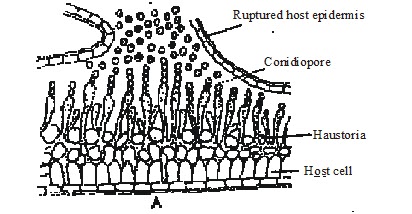
Cystopus - (A) V. S. of Infected leaf of Brasslca showing conidia ( B ) Conidiophore 2. Pythium
Q.3. Give the characteristics and life cycles of the Pythium.
Related Questions -
Q. Describe the life cycle of Phythium with the help of suitable diagrams. (2013)
Q. Describe the systematic position and important characters of Pythium. (2013)
Ans. Sytematic Position: -
Division - Mycota
Sub-division - Eumycotina
Class - oomycetes
order - peronosporales
Family - Pythiaceae
Genus - Pythium

1. Characters: -
1. Pythium belong to family Pythiaceae.
2. Family Pythiaceae includes both aquatic and terrestrial species, some are amphibious.
3. The members of this family exhibits the tendency in adaptation of land habit. Few truly aquatic species parasite algae and small protozoa.
4. Many of the terrestrial forms parasites or crop plants and cause serious diseases. Other are saprophytic. Some of the saprophytic species can live as facultative parasites and the parasitic species exhibit saprophytic tendencies. The mycelium is extensive1y developed and consists of branched, coenocytic hyphae.
5. Coenocytic hyphae do not show any distinction into rhizoidal and aerial hyphae.
6. The septa appear only to wall of the injured parts or to separate the reproductive cells.
7. These septa are made of solid plates and have no pore. Th hyphae is most of the species are intrace1lular ( penetrate the host cells) and usually of regular diameter. The haustoria are produced in some species.
2. Reproduction: -
(i) Asexual Reproduction: -
Asexual Reproduction in Pythium takes place by means of zoospores which are produced in small, globular or oval, sac-like sporangia. The sporangia are formed singly and terminally at the ends of somatic hyphae which project into the damp atmoshpere from the mycelium within the host tissue. There are no specialized sporangiophores. The sporangia measure 15-26 microns in diameter. Sometimes the sporangia are intercalary. The high humidity in the air promotes the growth of the fungal mycelium and the production of zoosporea.


Fig. Pythium sp.stages in asexual reproduction.
Each sporangium is a multinucleate structure. It is separated from the rest of the hypha, which still retains the power of elongation, by means of a transverse septum. The sporangium is pushed aside and the hyphal tip grows to form the second sporangium. The sporangium is filled with hyaline cytoplasm containing numerous nuclei.
2. Sexual Reproduction: -
Sexual reproduction in Phythium is oogamous type occurs by gametangial contact at the end of the growing season. At this time the food is nearly exhausted. There is not enough moisture for growth. At this stage the host has been killed and the fungus is living saprophytically. The sex organs are formed within the dead tissues of the host. The male sex organ is called antheridium and the female oogonium. They are developed in close proximity on separate short, lateral hyphae arising from the same mycelium Pythium debaryanum is thus Homothallic.
Development of Oogonium: -
The tip of the female hyphae inflates to from a globular swelling. Hyphal cytoplasmic contents migrate into the terminal swelling. Ultimately the terminal swelling is cut off from the parent hyphae by a basal hyphal plug or septum usually after but often before antheridial contact. The separated terminal swelling usually functions as the young oogonium.
Ultrastructure of Young Oogonium: -
Young oogonium. has a thin wall. Within the oogonial wall is the plasma membrane enclosing an e1ectron-dense cytoplasm rich in endoplasmic reticulum and ribosomes. Randomly packed in the cytoplasm are vacuoles and vesicles of various types. Some of these contain a dense spherical storage body and are called the reserve vesicles. Lipid Vesicles are small.
Structure of Mature Oogonium: -
At the time of fertilisation the oogonial contents undergo considerable reorganizaton. The organelles, vacuoles and vesicles move to their destination. The mitochondria, all the nuclei except one and vacuoles migrate to the periphery, the reserve vesicles and lipid vesicles move inwards and accumu1ate in the central area of the oogonium. Infact when the fertilization tube penetrates the mature oogonium its protoplast is differentiated into two zone, outer periplasm and inner ooplasm.
1. Periplasm: -
It is the narrow outer zone living the oogonial wall. It has spongy, or vacuolate protoplast. Apparently it seems to have been formed by shrinking away oogonial protoplast from the wall, but it is in fact formed by the coalescence and deterioration of vacuoles. Much of the lamellar endoplasmic reticulum accumulates around the periphery of the central dense area forming a layer parallel to the oogonial wall. The periplasm present at the outside of this layer contains surplus nuclei, mitochondria along with other organelles.
2. Ooplasm: -
The large central portion of the oogonium filled with granular electron dense cytoplasm constitutes the ooplasm; provided with a single nucleus and food reserve in the lipid and reserve vesicles. The uninucleate ooplasm functions as an oosphere or egg.


Pythium sp. Various stages in the development of sex organs and fertilization.
Development of Antheridium: -
Antheridium develops terminally on a short male or antheridial branch arising laterally either from the oogonial stalk or a neighbouring hypha. During early development antheridial branch curves towards the oogonium. It than comes in contact with the oogonial wall and flattens against it with the tip getting slightly inflated. The hyphal contents migrate into the enlarged tip. It is eventually separated from parent hypha by a hyphal plug to function as an antheridium.
The antheridium in.Pythium is hence applied to the side of the oogonium and is called paragynous.
Structure of Antheridium: -
It is an elongated, club-shaped structure much smaller in size than the oogonium. It has a thin wall, inner plasma membrane which in the young stage encloses an electron dense cytoplasm. The cell organelles (ribosomes, endoplasmic reticulum, nuclei, mitochondria and dictyosomes) are randomly packed in the cytoplasm. At muturity the antheridial protoplast becomes differentiated into a central unnucleated portion which functions as the male gamete and the outer periplasm. The surplus nuclei and other organelles migrate into the periplasm where they ultimately degenerate.
Fertilization: -
The gametangial contact occur at an early stage of development usually by the curvature of the male hypha, rarely by the female hypha. The tip of the antheridium applies itself closely to the oogonial wall and becomes flattened at the point of contact. The intervening walls of the mature sex organs dissolve at the point of contact. The flattened tip of the antheridium puts out a fine; tubular process known as, the fertilization tube or the conjugation tube. This tube penetrates the oogonial wall through the pore, pierces. the periplasm and dips into the ooplasm, it opens here and emits a single male nucleus together with a certain amount of cytoplasm. This cytoplasm mingles with cytoplasm of the egg. The male nucleus fuses with the female nucleus.
Post - ferti1ization Changes: -
Soon after fertilization, the egg secretes a thin membrane around it. A thick wall is subsequently deposited at this layer. The organelles involved in the synthesis and supply of material for this wall are reported to be the organelles and cytoplasm in the periplasmic space and the lamellar endoplasmic reticulum layer which delimits the periplasm from the ooplasm. With the formation of thick wall, all these structures disappear in the periplasm. Meanwhile the numerous small reserve vesicles in the developing fertilized egg enlarge and coalesce to form a single, large, dense globule in the centre. It is surrounded by the rapidly enlarging and closely packed lipid globules of various sizes. The resultant thick-walled structure containing reserve food material and scanty cytoplasm is the mature oospore.
Fine Structure of Oospore: -
Mature oospore is loosely surrounded by the thin oogonial wall. The periplasm having disappeared by now there is empty space between the oogonial wall and the oospore. The oospore thus partially fills the oogonial cavity and is described as aplerotie. The mature oospore has a thick smooth wall. The oospore wall is differentiated into two layer , the outer exine and the inner wall intine. The exine is opaque. The intine is comparatively thicker and electron transparent. The thick oospore wall contain centrally located large, dense spherical area surrounded by closely packed lipid globules of various sizes filling the space between the oospore wall and the central area. The scanty cytoplasm forms a thin layer around the central structure and lies between the lipid vesicles. This central area of the oospore has been differently interpreted by various investigators. Mckeen (1975) terimed this central area of the oospore as the reserve globule formed by the fusion of the reverse vesicles. The oospore is a resting structure. The nature of thick oospore wall provides protection and the large amount of lipid globules furnishes energy needed for its long dormancy.
After fertilization, the antheridium adhering to the oogonial wall reatains its characteristics shape as an empty or nearly empty shell the unfertilised egg behave like an oospore and is known as parthenospore.

Fig. stages in the germination of oospore and differentiation of zoospores.
During favourable conditions the oospore germinates. The diploid oospore nucleus undergoes repeated divisions. The increase in the. number of nuclei is accompanied by increase in the number of mitochondria The central globule begins to disintegrate and becomes surrounded by vacuoles indicating its digestion and absorption. Simultaneously the middle layer is eroded and middle layer of the oospore wall is pitted and becomes thin in the region of germ tube emergence. The emerging germ tube with its wall continuous with the inner layer of the oospore wall pushes through the middle and outer layers, at this site. After emergence from the oospore wall the germ tube presses against the oogonial wall and finally breaks through it one of the following ways.
(1) It grows into a small hypha. The latter infects the host and forms the starting point of the fresh mycelium.
(2) The tip of the hypha swells to form a vesicle like sporangium. The entire oospore protoplast migrates into the latter. The terminal sporangium may either behave like a zoosporangium and produce the zoospores or gets detached. The detached sporangium behaves like a condium and germinates directly by putting out a germ tube.
(3) In this mode of germination e.g. Phythium anandrum and Pythium mamillatum the contents of the oospore divide to form biflagellate zoospores which are emptied into a vesicles. The vesicle soon disappears and the zoospores are liberated. Each librated zoospore germinates and infects the host in the usual manner and forms a new fungus mycelium.
3. Pythium Life Cycle: -

Fig. word diagram of life cycle with zygoric meiosis.
The diploid nucleus of the oospore prior to germination divides a number of times. According to Edison (1915 ) the first two successive divisions are meiotic. On the basis of zygotic meiosis scheme the vegetative thallus (mycelium) or pythium is considered haploid. It bears haploid gametangia containing male and female gametes towards the end of the growing season. The male and female gametes fuse to form the oospore which is the only diploid structure representing the saprophyte in the life cycle of Pythium. All other structures, the zoospores, mycelium, gametangia and gametes represent thes gametophyte such a life cycle with a prolonged haploid vegetative phase (haplo-phase) and a single-celled diploid oospore representing the diplophase is called haplontic.

Fig. Word diagram of life cycle with gametangial meiosis.
It is characterized by zygotic meiosis and haploid adult ( mycelium ), Sansome (1961 and 19 63) working on Pythium debaryanum discorded the scheme of zygotic meiosis advance evidence indicating that Pythium has a diploid vegetative thallus (mycelium ) and gametangia (oogonium and antheridium ) are meiotic. Dennet and Stanghellini (1977) have also advanced genetic and cytological evidence for diploid life cycle and gametangial meiosis in Pythium aphanidermatum. On the basis of gametangial scheme of meiosis Pythium has a prolonged diploid vegetative phase represented by the zoospores , mycelium and gametangia ( oogonia and antheridia ). The haploid phase is extremely reduced. It is represented only by the gametes ( male nucleus and oosphere ).
This type of life cycle is called Diplontic. It is characterised and diploid adult ( mycelium ). The vegetative phase whether diploid or haploid is prolonged by the formation of sporangia which germinate directly or indirectly by the zoospores during growing seasons.
